
Newborn fontanelles: what are they and what are they for?
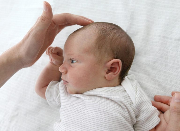
From the first visit, the pediatrician checks the condition of the newborn's fontanelles .
Haven't you ever seen him gently touching some points on the little one's head?
Knowing how to locate and evaluate the cranial fontanelles is very important for every parent to immediately check the condition of the child in case of abnormalities.
Find out what the newborn's cranial fontanelles are, how many there are and what their purpose is by reading this article.
Before starting, I'll leave you some useful information:
- If you would like to schedule a studio visit with me, you can find me here.
- You can also find me on Instagram with the profile @drsilva.com_official
- In the video courses section you will find how to promote the psychomotor development of the newborn.
Fontanelles of the newborn: introduction to the anatomy of the skull

Before seeing what the fontanelles of the newborn are, I will make an introduction to the anatomy of the skull . In fact, in this way it will be easier to understand the position of the fontanelles and their function.
Formation of the skull
The human skull is made up of 22 bones , some of which are even and symmetrical between the right and left sides.
The main function of the skull is to protect the noble parts of the head :
- Brain;
- Cerebellum;
- Brainstem;
- Sensory organs.
What differentiates the skull of the child/adult from that of the newborn is the presence of fontanelles. Instead, what they have in common are the number of bones and the sutures. We will talk about the latter in the next chapter.
The skull can be divided into two:
- Splanchnocranium : Composed of 14 bones at the front of the head. Provides space for the functioning of the main sense organs.
- Neurocranium : composed of 8 bones and provides space for the organs of the central nervous system (cerebrum, cerebellum and brainstem).


Cranial sutures of the newborn
We have seen that the brain is made up of bones and sutures.
What are these last ones?
The cranial sutures are the joints between one bone and another and are characterized by dense fibrillar connective tissue .
Let's see together the main ones that also concern the cranial anatomy of the newborn:
- Metopic suture : centrally joins the two frontal bones from the nose to the anterior fontanel. This suture is destined to fuse by the time the child is 5 years old. If ossified from birth it causes trigonocephaly, or a pointed shape of the head.

- Coronal suture : joins the frontal bone and the parietal bone on both the right and left.

- Sagittal suture : joins the two parietal bones along the meridian line of the skull, starting from the anterior fontanelle to the posterior one.

- Lambdoid suture : joins the occipital bone and the parietal bone on both the right and left.

What are the newborn's fontanelles?
Surely you have heard at least once the term fontanel of the newborn in reference to that soft part in the skull of the little one that seems to pulsate at times. In fact, for this reason it is called fontanel.
These are soft and membranous parts where at least 2 cranial sutures or 3 skull bones cross. They are characterized by a fibro-cartilaginous tissue and are 6.
Yes, you understood correctly! Everyone knows the anterior and posterior fontanelle, but few know that there are 4 other lateral ones, even and symmetrical.
But let's look at them in detail.
Bregma: anterior central cranial fontanelle
The anterior fontanelle of the newborn is diamond-shaped and varies in length from 1 to 3 cm.
The right and left coronal sutures and the sagittal suture (median cranial suture that forms between the two parietal bones) converge here.
Lambda: posterior central cranial fontanelle
The posterior fontanelle is triangular in shape and measures approximately 0.5 cm in size.
It is located behind the skull and the sagittal suture and the lambdoid suture join here.
Sphenoid: anterolateral cranial fontanelle
This small fontanelle is positioned between the sphenoid bone, the parietal bone, the temporal bone and the frontal bone.
Being symmetrical, it is found on both the right and left side.
Mastoidea: Posterolateral cranial fontanelle
This second small fontanelle is located just behind the ear between the temporal bone, the occipital bone and the parietal bone.
This one, like the previous one, is located symmetrically on the right and left side.
Closure of the newborn's fontanelles
The newborn's fontanelles are destined to close at different times:
- Bregma , the anterior one, ossifies around the 18th month of life and within the second year. According to statistics, it is closed in 1% of children at the 3rd month, in 38% at the 12th month and in 96% at the 24th month;
- The posterior fontanelle tends to close in the first two/three months of the baby's life;
- The sphenoid bone ossifies around 6 months ;
- The mastoid , on the other hand, between 6 and 18 months .
As we said, the closure occurs through a process of ossification .
What does it mean? In ossification, phosphorus and calcium salts are deposited on the fibrous tissue and transform it into bone tissue. For this to happen, there must be an adequate amount of these salts, a balance in their metabolism and a correct dose of vitamin D.

Closing too fast
The closure of the anterior fontanelle is said to be precocious if it closes within 4 months of the newborn's birth .
In this case, a careful evaluation by the pediatrician is necessary to understand if there is a risk of two problems:
- Microcephaly : is the reduction of the circumference below the norm and is often a symptom of anomalies in the brain structures;
- Craniosynostosis : is the pathology that causes an early closure of the fontanel, resulting in an abnormal development of the head.
Delayed ossification
In contrast to premature closure, there is delayed ossification of the fontanelle in the newborn .
This case study could also indicate problems in the child:
- Thyroid problems ;
- Rickets : a pathology linked to a low quantity of vitamin D;
- Achondroplasia : disease related to the growth plate;
- Osteogenesis imperfecta : pathology characterized by bone fragility;
- Trisomy 21 (Down syndrome).
What are they for?
If fontanelles are present only in newborns and are destined to disappear later, what purpose are they?
These allow the skull to shape so that the head can emerge from the birth canal .
Later, however, I allow the growth of the brain and the bones of the skull . Just think of the growth of the circumference of the newborn which goes from about 35 cm at birth to 47 cm around the year.
These are the functions of the fontanelles, but they can also be a signal about the health of the little one.

Sunken fountain
If the anterior fontanelle is markedly depressed with respect to the plane of the cranial bones, it may indicate a state of dehydration in the newborn.
It can happen in case of intestinal flu in the little one.
Curved fountain
When the fontanelle is markedly raised and associated with an altered state of consciousness and reactivity of the newborn, it is possible that there is an increase in intracranial pressure . This, associated with fever and grayish color or red patches on the skin, may indicate meningitis . It is absolutely important to go to the emergency room immediately to exclude this possibility.

As you may have understood, the fontanelles make the head easily moldable . This is helpful during birth and for brain growth, but it is also the reason why the newborn can experience plagiocephaly or other positional cranial deformations . In fact, if the baby always keeps his head positioned on the same side, the pressure exerted on that side causes the skull to undergo an alteration of shape.
For this reason, it is important to know and put into practice all the necessary precautions to prevent or promptly treat flat head. You can find all this information in my video course “ Bye bye flat head ” and in “ How to Treat Flat Head and Positional Plagiocephaly in Newborns ”.